


Bovine Viral Diarrhea Virus (BVDV)
Description: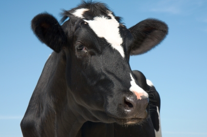
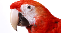
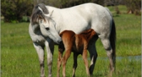
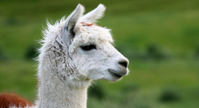
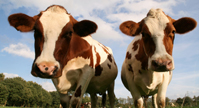
Bovine Viral Diarrhea Virus (BVDV) is a Pestivirus classified into two genotypes, "Type 1" and "Type 2," that infects domestic and wild ruminant herds world-wide. In addition to cattle, BVDV is also known to infect camelids and deer. Related viruses include hog cholera virus in swine and border disease in sheep. For cattle producers, the virus causes an estimated $2 billion per year in economic losses through decreased weight gains, decreased milk production, reproductive losses, and death.
Within each genotype there are hundreds of BVD virus types (both cytopathic and non-cytopathic), each of which differ significantly in their ability to cause disease.
Two types of infection occur in cattle: Transient (acute) Infection (called TI) and Persistent (chronic) Infection (called PI). TI infections are short-term (weeks). Acute BVD virus infection is generally acquired after birth and TI cattle become immune and clear of BVD virus. Of the BVD infection in cattle, currently 95% of BVD infections are TI. Persistent (chronic) Infections (PI) are life-long infections acquired in utero. This means that only fetal infections generally result in BVD-PI. Around 5% of BVD infections in cattle are PI. PI BVD cattle never become immune and are the major source of virus spread in herd. Surprisingly, more than 90% of BVD-PI calves are born from normal dams (no prior BVDV exposure).
The BVD virus can cause respiratory disease, enteritis, stillbirth, abortion, and mucosal disease. In utero, BVDV infection can induce intolerance, causing animals to be persistently infected (PI) for life. PI animals shed several billion viral particles a day and serve as reservoirs of BVDV in a herd.
Transmission of BVDV from one animal to another typically occurs via the oral/nasal route. Acute or persistently infected (PI) cattle shed over 2 billion viral particles everyday in mucus, saliva, feces, and urine. Risk of BVDV transmitted, to a much lesser extent, is possible by contaminated equipment, semen, fetal calf serum, and biting insects.
The primary source of new BVD infections within the herd are PI animals. Fetuses infected at less than 150 days gestation suffer high death rates, birth defects, or become persistently infected. Calves infected after 150 days gestation, can develop a sufficient antibody response to the BVD virus and potentially eliminate the infection. All calves born to PI dams will become PI BVD animals.
BVD Disease in Cows:
Many factors influence the severity of BVD in cattle including the viral type, reproductive status, immune status, stress, presence of additional pathogens and the age of the animal. As with most immunosuppressive diseases, BVD infections decrease the immune system's capacity for fighting infection by reducing the white blood cell count. Much of this is due to suppression of the macrophages, neutrophils and lymphocytes. These cells are part of the immune system's responce to infection. BVD virus can localize in tissues and remain present for the life of the animal.
In cattle with clinical disease, BVD virus is present in the blood from about day 3 to 11 after the virus has entered the animal. Clinical signs start as early as day 9 to 13 after the initial infection. Three types of clinical disease are recognized: 1) Mucosal disease, where both cytopathic and non-cytopathic virus is present; 2) severe acute BVD; and 3) hemorrhagic syndrome. Along with clinical disease there is also non-clinical or inapparent disease.
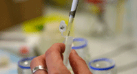
USDA-licensed Test for BVDV:
In 2010, Life Technologies(TM) Corporation introduced the first United States Department of Agriculture (USDA)-licensed real-time PCR (rtPCR) test kit to detect BVDV, in cattle, camelids, and swine . The new rtPCR kit for BVDV detection provides a reliable tool to test for the presence of Type 1 and Type 2 BVDV. The reaction also demonstrated consistent results when applied to sample containing 18 BVDV-1 subgenotypes and 6 BVDV-2 subgenotypes. All subtypes were detected, further demonstrating the assay's diagnostic sensitivity.
Sample Collection Instructions:
Ear Notch: A medium pig ear-notcher or ear-notching tool that yields a 1 cm notch works best if used on the lower aspect of the ear where the notch can be seen from a distance yet does not affect the function of the ear flap.
Clean the notching tool with disinfectant, then ALWAYS clean away any disinfectant with distilled water. It is important to remember that any residual disinfectant on the notching tool can cause false-negative results, therefore thorough rinsing with clean water is essential. Place the notch into a labeled, dry, red top collection tube with no formalin or other liquids. Collected ear notch MUST be free of contamination including dirt, feces, tattoo ink or BVD vaccine.
Store collected ear notches for a maximum of 72 hours at refrigerator temperatures and ship over.
Prevention and Control:
Studies show that colostrum containing BVDV antibodies may help protect a calf from BVDV Types 1 and 2. A dam with BVDV antibodies will transfer temporary passive immunity to the calf through her colostrum, providing a level of protection that usually lasts 3-6 months. Critical periods for new BVDV infections are in calves 3-6 months of age when colostral titers are gone, and at 9-10 months when ovaries and testicles are becoming active.
The groups most susceptible to BVDV are seronegative animals, calves that did not ingest sufficient colostrum, and cattle under environmental- or disease-related stress.
The primary source of infection are PI cattle as they continuously shed large amounts of virus in their feces, saliva, and nasal discharge. Testing allows producers to identify PI animals and cull them to decrease the amount of BVD virus circulating on the dairy.
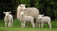
Another source of BVD exposure is direct or indirect contact with infected cattle in neighboring pastures. Producers should be aware that sheep, goats, and deer also carry BVDV and can cross-infect cattle. These cross infections seldom cause visible disease in cattle.
It is important to remember that testing is only one component of a complete BVD control program. Proper vaccination, biosecurity and biocontainment are important issues to also address when constructing a comprehensive managment plan to deal with BVDV.
References:
Multiple diagnostic tests to identify cattle with Bovine viral diarrhea virus and duration of positive test results in persistently infected cattle. Fulton RW, Hessman BE, Ridpath JF, Johnson BJ, Burge LJ, Kapil S, Braziel B, Kautz K, Reck A.Can J Vet Res. 2009 Apr;73(2):117-24.
Prevalence of bovine viral diarrhea virus infections in alpacas in the United States. Topliff CL, Smith DR, Clowser SL, Steffen DJ, Henningson JN, Brodersen BW, Bedenice D, Callan RJ, Reggiardo C, Kurth KL, Kelling CL. J Am Vet Med Assoc. 2009 Feb 15;234(4):519-29.
Submit a Sample for Testing:
To submit a sample for testing please go to test now.
To
order a sample collection kit please go to order sample collection kits.